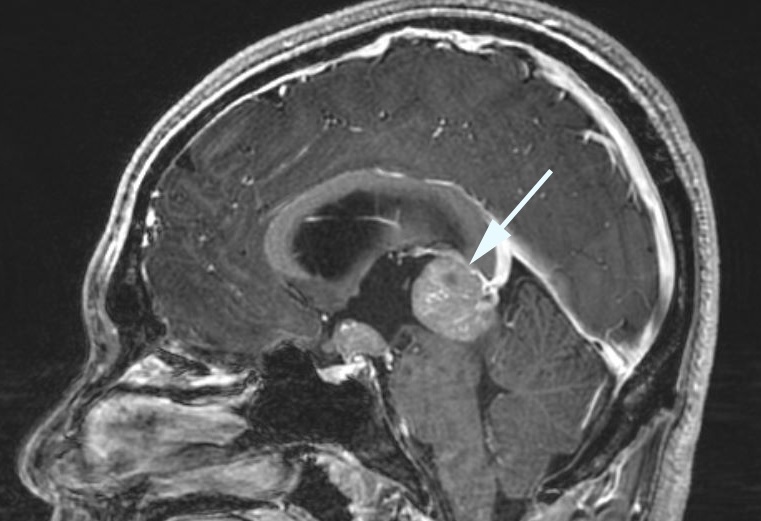
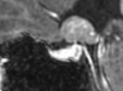
Suprasellar Germinoma
-
Suprasellar tumor arising from germ cells
-
Vision loss is caused by compression
-
Teenagers are most at risk
-
No sex predominance
-
Often cured by chemotherapy and radiation therapy
-
Core clinical features
-
Subacute or chronic vision loss
-
Hemianopic visual field defects
-
Optic discs appear normal or pale
-
Afferent pupil defect
-
Possible accompanying clinical features
-
Hypopituitarism, especially diabetes insipidus
-
Dorsal midbrain syndrome from a pineal region germinoma
-
Imaging features
-
Suprasellar mass that enhances avidly
-
Pineal mass often also present
-
Tumor deposits in cauda equina sometimes present (“dropped metastases”)
-
Other masses in sellar region
-
Arachnoid cyst
-
Epidermoid cyst
-
Langerhans cell histiocytosis
-
Sphenoid sinusitis or mucocele
-
Lymphocytic hypophysitis
-
Pituitary abscess
-
Multiple sclerosis
-
Neuromyelitis optica
-
Sarcoidosis
-
Spine MRI will be performed to exclude dropped metastases
-
Lumbar puncture will be performed to detect markers of nongerminomatous germ cell tumors
-
Biopsy of the lesion will be performed if imaging is not distinctive
-
Platinum-based chemotherapy is sometimes followed by low-dose brain radiation therapy
-
Permanent cure will occur in >90% with minimal or no lingering neurologic deficits
-
Visual recovery depends on how much damage has occurred before treatment began