Hypoplastic Optic Neuropathy
- Congenitally small optic disc, optic nerve, and optic chiasm
- Often accompanied by other forebrain dysgeneses
- Rare binocular variant called “superior segmental optic disc hypoplasia” (“topless optic discs”) associated with maternal diabetes
- Visual impairment caused by deficient optic nerve axons
-
Clinical features
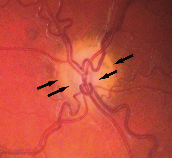
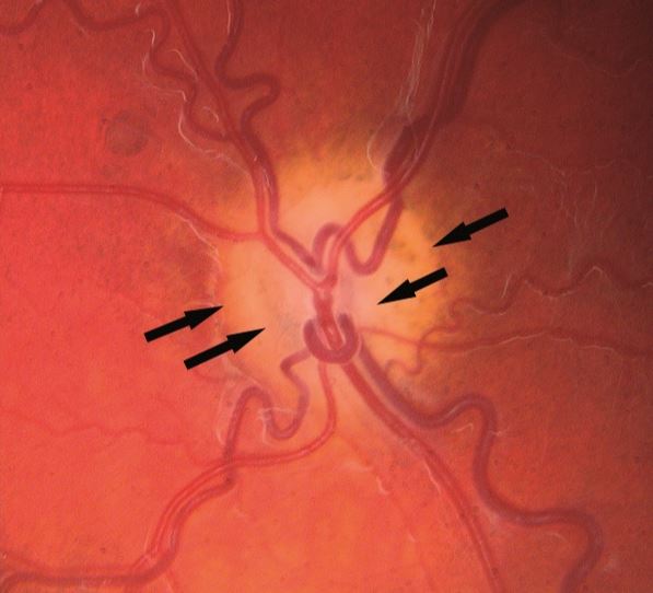
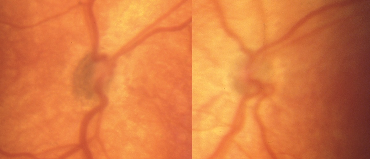
- Small optic disc diameter in one eye or both eyes
-
Trap: may be diagnostically challenging because it is hard to get a good ophthalmoscopic view in infants and hard to distinguish the true margin of the optic disc
- Inner pigment ring surrounding the margin of the optic disc and an outer pigment ring surrounding peripheral chorioretinal atrophy (“double ring sign”)
- Variant called “superior segmental optic disc hypoplasia” lacks superior optic disc structure (“topless optic discs”) and has inferior nerve fiber bundle visual field defects but no brain anomalies

-
Possible imaging features
- Small-caliber optic nerve
- Absent septum pellucidum and hypoplastic optic nerves and chiasm (de Morsier syndrome)
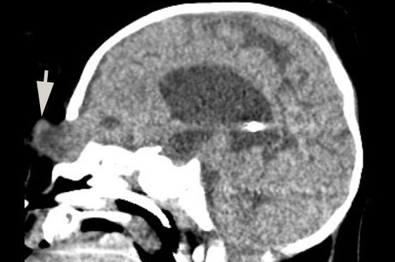
- Frontal encephalocoele
- Absent or displaced posterior pituitary bright spot

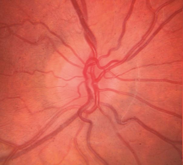


- Normal optic disc
- Perform brain MRI, looking for a small-caliber optic nerve and other forebrain abnormalities
- Look for ophthalmoscopic signs of optic disc hypoplasia if other forebrain or pituitary abnormalities have already been detected
- Anticipate deficient growth hormone and adrenocorticotropic hormone, especially if the pre-contrast T1 MRI sequence shows that the “posterior pituitary bright spot” is absent or upwardly displaced
- Alert the pediatrician that hypopituitarism places the child at risk of sudden death in a febrile illness
- Expect poor vision in some patients with optic disc hypoplasia, but…
-
Trap: the correlation between optic disc size and vision is weak, so do not issue predictions about visual potential until you can assess vision adequately when the child is older
- If you find superior optic disc hypoplasia, look for corresponding inferior nerve fiber bundle defects, and inquire if the patient’s mother was an insulin-dependent diabetic at the patient’s birth
- Visual dysfunction will remain stable
- Forebrain anomalies are more likely if hypoplasia affects both eyes
- Hormone deficiency is more likely if brain imaging shows forebrain anomalies