Non-arteritic Ischemic Optic Neuropathy
- Infarction of the prelaminar optic disc that causes acute vision loss
- Commonest explanation for acute optic neuropathy in middle-aged and elderly adults
- Probably a small-vessel ischemic stroke associated with standard risk factors for arteriolar sclerosis
- May also be precipitated by low systemic blood pressure, especially during sleeping hours, and exposure to interferon alpha-2b or other chemotherapeutic agents
-
Core features
- Acute, painless vision loss in one eye that may worsen over a maximum of 14 days
- Reduced visual acuity and/or nerve fiber bundle visual field defect in the affected eye
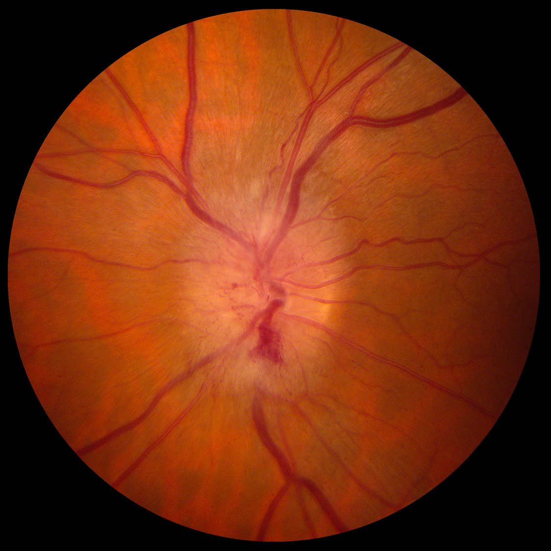
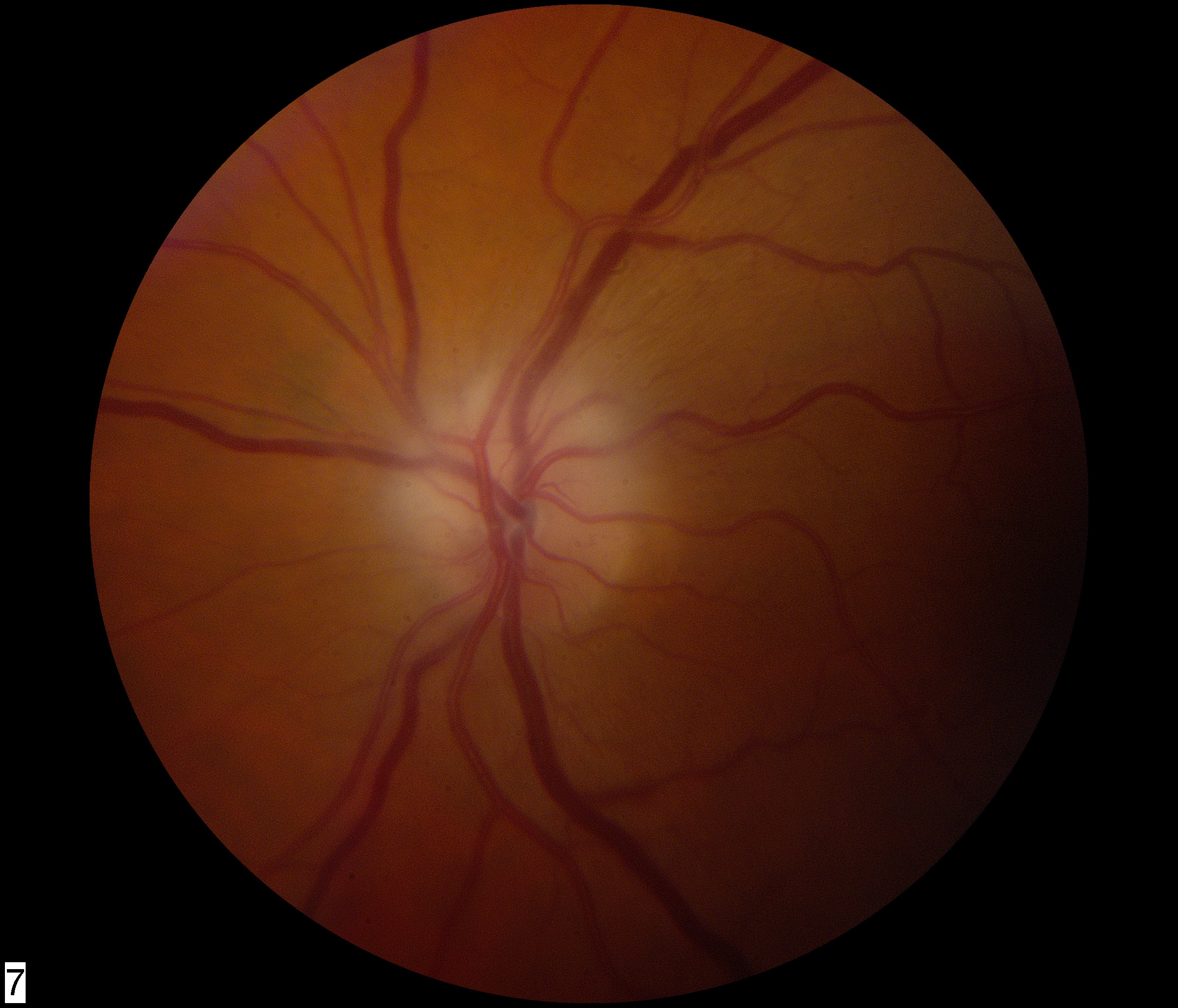
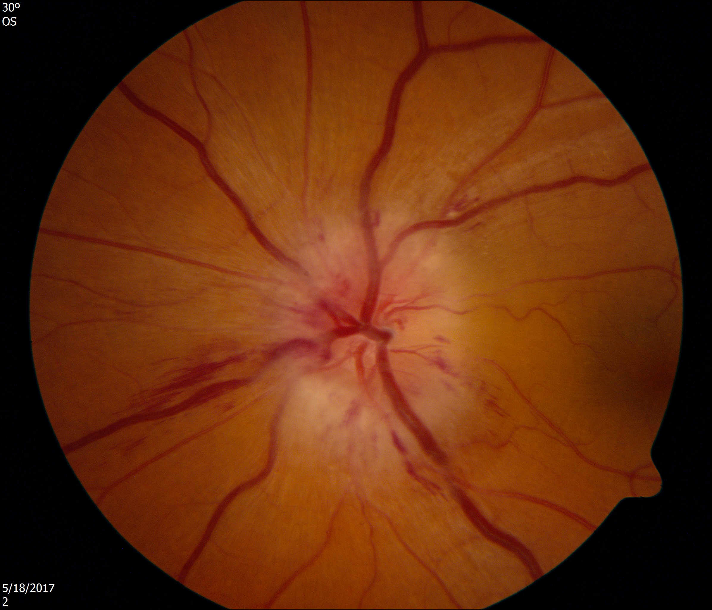
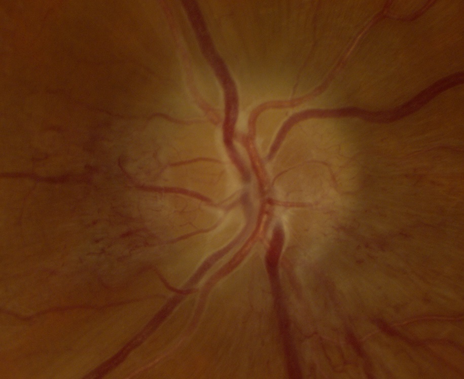
- Diffuse or segmental hyperemic or pallid edema of the optic disc elevation in the affected eye, sometimes sprinkled with hemorrhage
- Standard arteriolar sclerosis risk factors are usually present (systemic hypertension, diabetes mellitus, obesity, cigarette smoking, dyslipidemia, family history of premature heart attack or stroke)
- No symptoms of polymyalgia rheumatica or giant cell arteritis
-
Possible accompanying features
- Small or absent optic disc physiologic cup in the unaffected eye (“disc at risk”)
Trap:
may be difficult to distinguish from arteritic ischemic optic neuropathy, papillitis, infiltrative (neoplastic) optic neuropathy, and other causes of acquired optic disc elevation
- Papillitis
- Arteritic anterior ischemic optic neuropathy
- Infiltrative (neoplastic) optic neuropathy
- Diabetic papillopathy
- Leber hereditary optic neuropathy
- Hypertensive optic disc edema
- Paraneoplastic optic neuropathy
- Papilledema
-
Rule out arteritic ischemic optic neuropathy in patients aged more than 55 years by excluding new
- Jaw claudication
- Scalp tenderness
- Limb girdle aches
- Anorexia
- Fever
- Elevated sedimentation rate, C-reactive protein, platelet count
- Perform temporal artery biopsy if you cannot exclude arteritic ischemic optic neuropathy
- Reduce discretionary arteriosclerotic risk factors with conventional measures and advise patients to avoid low blood pressure and agents used for erectile dysfunction because they divert blood from the optic nerves
- Discuss discontinuation of amiodarone because some authorities consider it a risk factor despite weak evidence
-
Tip: carotid imaging studies are not necessary because this condition is not caused by disease of large-caliber vessels
-
Trap: even if carotid imaging shows flow-limiting cervical carotid stenosis, ischemic optic neuropathy should not be used as a justification for carotid endarterectomy or stenting
- Vision loss in the affected eye may worsen within 2 weeks of onset
- Vision recovery is negligible
- Recurrence in the affected eye is very rare
- Fellow eye has a 15%-20% chance of having a similar event within 15 years
- No evidence that conventional stroke preventive measures reduce the risk of second eye involvement
- Risk of non-optic ischemic stroke is probably higher than in a non-NAION risk-matched population, but the evidence is tentative
-
Trap: adult patients who have acute unilateral vision loss, a swollen optic disc, and an afferent pupil defect, will often receive a mistaken diagnosis of optic neuritis (papillitis). When brain MRI discloses multiple high signal spots in the cerebral white matter on T2 and FLAIR sequences, such patients will be labeled as having multiple sclerosis. But those scattered white spots reflect the ischemic demyelination of systemic hypertension or diabetes, rather than the autoimmune demyelination of multiple sclerosis. Although people will say that the location of the ischemic white spots and autoimmune white spots is different, there is much overlap. Treatment with corticosteroids and chronic immune-modulating agents is not indicated. The appropriate management consists of reducing the factors that might predispose to a similar ischemic event in the unaffected eye!
-
Tip: non-ophthalmic physicians are generally unaware that low blood pressure may precipitate this condition and may resist advice to avoid a relatively low blood pressure regimen, which is beneficial to heart and kidney function