Visual Cortex Lesions

- Focal damage to the visual cortex
- Common cause: stroke
- Uncommon causes: neoplasm, vascular malformation, hemorrhage, posterior reversible encephalopathy syndrome (PRES), Alzheimer disease, encephalitis
- Homonymous hemianopic visual field defects and rarely a monocular temporal crescent defect
-
Possible accompanying features
- Ipsilateral brow pain because of referred pain from irritated occipital meninges
- Hallucination of flickering lights or shapes (“scintillations”) in the hemifield contralateral to the lesion
- Hallucination of recently viewed objects (“palinopsia”)
- Illusion of grossly distorted perception of viewed objects (“cerebral metamorphopsia”)
- Illusion of reduplicated viewed objects (“cerebral polyopia”)
- Illusion that moving objects have comet-like trails
- Illusion that moving objects are not moving (“akinetopsia”)
- Illusion that moving objects are broken up into consecutive frames
- Impaired reading but intact writing and spelling (“alexia without agraphia”) with left hemisphere lesions
- Impaired recognition of familiar objects with bilateral lesions
- Impaired recognition of familiar and famous faces (“prosopagnosia”) with bilateral lesions


-
Brain MRI usually shows the lesion, but it may be subtle
or even inapparent in the following conditions
- Recent severe systemic hypotension causing hypoxic-ischemic brain injury
- Acute migraine
- Focal status epilepticus
- Alzheimer disease (“visual variant,” “posterior cortical atrophy”)
- Nonketotic hyperglycemia
- Creutzfeldt-Jakob disease (“Heidehain variant”)
- Focal encephalitis (“cerebritis”)


- Symptoms may be poorly expressed by the patient or dismissed by the caregivers as psychogenic because visual acuity and structural aspects of the eye examination are normal
- Be alert for distinctive visual and accompanying symptoms
-
Look for the following features on visual field examination to help localize the lesion
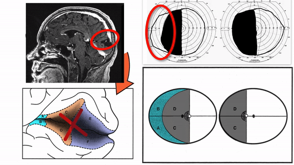
- Homonymous quadrantanopia (defects confined to the upper or lower quadrant, with borders aligned to vertical and horizontal meridians), which suggests a lesion of the posterior optic radiations or visual cortex
- Homonymous paracentral scotomas (small defects adjacent to one side of fixation), which suggest a lesion of the posterior visual cortex
- Macular-sparing homonymous hemianopia (defects sparing central 5 to10 degrees of visual field), which suggests a lesion that spares the posterior visual cortex
- Monocular temporal crescent scotoma (defect affecting unpaired peripheral temporal field in one eye), which suggests a lesion of the anterior visual cortex
- Temporal crescent-sparing homonymous hemianopia (defects sparing unpaired peripheral temporal field of one eye), which suggests a lesion that spares the anterior visual cortex
- Bilateral homonymous hemianopias (defects affecting both sides of the visual field), which suggest lesions of bilateral visual cortex


- Expect to find a correlative MRI abnormality, although it might be subtle or inapparent in the 7 conditions described in What does it look like?
-
Tip: watch out for ipsilateral brow pain as a symptom of occipital lobe infarction; the pain is referred from irritation of posterior meningeal trigeminal receptors
-
Tip: watch out for pure alexia with right homonymous hemianopias, and watch out for visual spatial disorders (including hemispatial neglect) with left homonymous hemianopias
- Depends on the nature of the underlying disorder
-
Tip: in acute stroke, tumor, encephalitis, or posterior reversible encephalopathy syndrome (PRES), visual field loss may be caused or exacerbated by superimposed non-convulsive status epilepticus, which is often accompanied by contraversive head and gaze deviation; rule out this condition with electroencephalography, and treat with anti-convulsive agents
- Patients with extensive unilateral homonymous hemianopias and intact cognition may bump into objects in their blind hemifield, but they can conduct most activities of daily living except safe driving
- Patients with homonymous hemianopias that do not allow at least 60 continuous degrees on both sides of fixation along the horizontal meridian and 30 continuous degrees on both sides of fixation along the vertical meridian will not be permitted to drive in most jurisdictions
-
Trap: optical devices (spectacle-based mirrors, press-on prisms) that shift visual targets into the intact hemifield do not provide adequate vision for safe driving
-
Trap: formal vision training programs do not improve on patients’ spontaneous ability to learn to explore their blind hemifield