Lateral Geniculate Body Lesions
_Encephalitis_Ford.png)
- Focal damage to the lateral geniculate body
- Causes of unilateral lesions: neoplasm, aneurysm, vascular malformation, head trauma, neurosurgical procedures, stroke, inflammation
- Causes of bilateral lesions: systemic hypotension, vasculitis, encephalitis, rapid correction of hyponatremia (“osmotic demyelination,” “extrapontine myelinolysis”)
- Visual acuity is preserved with unilateral lesions but is often severely compromised with bilateral lesions
- With unilateral lesions, complete homonymous hemianopias are most common because the lesion typically destroys the entire lateral geniculate body
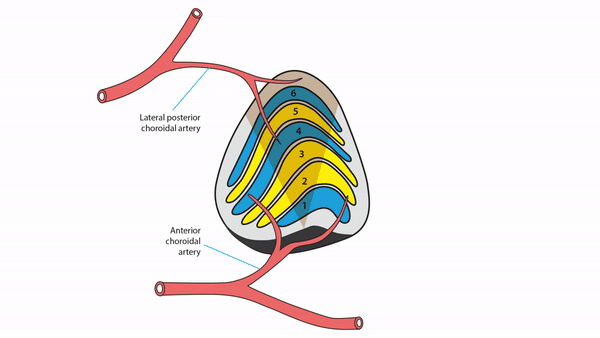
- With unilateral lesions, hourglass sectoranopia may occur following anterior choroidal artery occlusion
- With unilateral lesions, silhouette of hourglass sectoranopia many occur following lateral choroidal artery occlusion
- Ophthalmic manifestations may be the only clinical abnormalities
- Brain imaging may show the lesions, but the abnormalities are often subtle


_Encephalitis_arrows.jpg)
- Unilateral homonymous hemianopias from lesions elsewhere in the retrochiasmal visual pathway
- Perform visual fields on patients whose visual complaints are unexplained by ocular abnormalities
-
Tip: alert radiologists to the possibility of lateral geniculate body lesions in order to avoid overlooking subtle imaging abnormalities
- Treatment outcome depends on the cause