( of )
Correct: 0
Incorrect: 0
A 60 year old woman with diabetes mellitus is diagnosed with small cell lung cancer and a frontal lobe metastasis. She is treated with chemotherapy and whole brain radiation. Follow-up studies, including repeat brain MRI, consider her in remission. One year later, she develops acute vision loss in the right eye. There are no other new symptoms. Her diabetes is under control. The neuro-ophthalmic examination shows reduced visual acuity, a right afferent pupil defect, and a nerve fiber bundle defect in the right eye with an otherwise normal examination. The examination of the left eye is normal. Neurologic examination is normal. An axial post-contrast T1-weighted brain MRI scan looks like this.
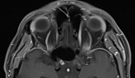
The diagnosis is
Incorrect
Correct!
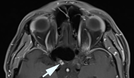
 This MRI is showing a discrete area of enhancement in the prechiasmatic (intracranial) right optic nerve.
Although such signal changes do occur here in optic neuritis, their location is typical for radiation-induced optic neuropathy. Moreover, the timing is
perfect for this condition, which is based on endothelial damage and secondary infarction.
Neither neoplastic nor paraneoplastic optic neuropathy shows such a discrete MRI signal abnormality. Paraneoplastic optic neuropathy is extremely rare and is usually part of a posterior uveitis.
Radiation-induced optic neuropathy is also a rare event when proper radiation protocols are conducted. But patients with underlying vasculopathy (neurofibromatosis, diabetes, hypertension) are at higher risk. Even those without these predisposing conditions are vulnerable if the cumulative x-ray dose exceeds 5,000 cGy. Proton beam radiation can also cause this optic neuropathy.
Although hyperbaric oxygen and anticoagulation treatments are often proposed, they have never been shown to be effective. Unfortunately, stepwise decline in vision often occurs, sometimes involving both optic nerves.
Incorrect