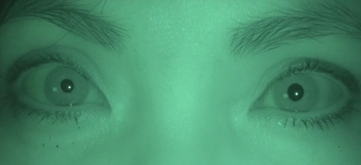
Pupil Examination
-
Step 1:
assess the size of the pupils in dim illumination
- Position your light below the patient’s chin and shine the light on the upper face, using the minimum amount of light needed to judge pupil size
- Measure pupil size in both eyes with the gauge on the Snellen near visual acuity card
-
Step 2:
assess the constriction of the pupils to direct light
- Position your light 3 inches below the patient’s right eye and shine the light directly on that eye; perform the same maneuver on the left eye
- Note the pupil size in each eye at maximal constriction to light
- If either pupil does not constrict normally to light, test for “light-near dissociation” by comparing pupil constriction to light and to a target held within 3 inches of the eye
-
Step 3:
hunt for a relative afferent pupil defect
- Shine your light first on the right pupil, then on the left pupil, moving the light across the nasal bridge
- Look for relatively reduced constriction or dilation of one pupil as the light is shined on it
-
Trap: a relative afferent pupil defect need not display dilation; a small relative afferent pupil defect may be diagnosed if there is a consistent difference in pupil dynamics on this test
-
Step 4:
if there is anisocoria and both pupils constrict normally to light, you must rule out a Horner syndrome by instilling apraclonidine 0.5% in each eye, even if there is no ptosis ipsilateral to the smaller pupil
- Look for reversal of anisocoria after 30 minutes
-
Trap: instilling apraclonidine in children aged 2 years or less may cause acute cardiovascular side effects, so use cocaine 10% instead
- Physiologic anisocoria: a pupil size difference of 1mm or less in dimmest illumination, both pupils constricting normally to light, and a negative topical apraclonidine or cocaine test
-
Pathologic anisocoria: there are 7 causes
- Horner syndrome: lesion in the oculosympathetic pathway that allows both pupils to constrict normally to light, but causes the affected pupil to dilate poorly in darkness; there is usually mild ipsilateral ptosis; the apraclonidine test causes reversal of anisocoria
- Third nerve palsy: lesion in the pre-ganglionic nerve segment that causes a dilated, poorly constricting pupil, along with ocular ductional deficits and/or ptosis of varying severity
- Dorsal midbrain syndrome: lesion in the midbrain tectum that causes large pupils that constrict poorly to light and better to a near target, and some combination of vertical gaze impairment, lid retraction, and clonic convergence retraction on attempted upgaze
- Tonic (Adie) pupil: ciliary ganglion or ciliary nerve lesion that causes slow segmental iris constriction to a near target, slow dilatation on refixation at distance, and pupil constriction to dilute pilocarpine; considered a limited dysautonomia
- Pharmacologic mydriasis: exposure to a topical anticholinergic agent that causes a mydriatic pupil that fails to constrict to pilocarpine at 0.5% concentration or higher; be aware that topical sympathomimetic agents can also cause a mild pharmacologic mydriasis, and that the affected pupils may not constrict normally to direct light
- Iris sphincter lesion: iris damage from eye surgery, trauma, uveitis, or dysplasia that causes the affected pupil to be irregular and to constrict poorly to light; slit lamp examination usually discloses iris pathology
-
Relative afferent pupil defect: there are 3 causes
- Ipsilateral optic neuropathy or relatively greater ipsilateral than contralateral optic neuropathy
- Ipsilateral extensive retinopathy or relatively greater ipsilateral than contralateral retinopathy
- Contralateral optic tract lesion
- Light-near dissociation: pupil that constricts poorly to direct light and more amply to a target fixated at 3-inch distance from the eye; there are 3 causes
