Color Vision Examination
- Assessment of color (chromatic) vision
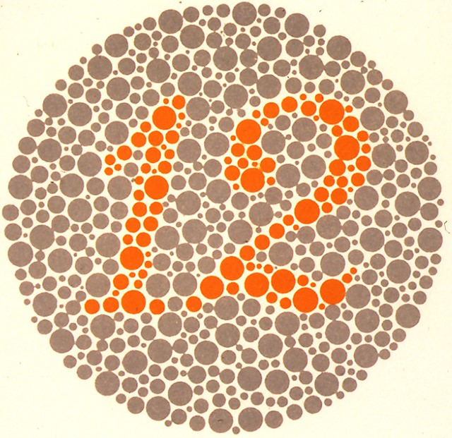
- Instruct the patient to identify items on the standard pseudoisochromatic color plate sets
- Score as a fraction-- correct answers over total plates presented
-
Trap: pseudoisochromatic plate testing is designed for congenital rather than acquired color vision disorders and tends to be insensitive in assessing acquired non-cone retinocortical lesions
-
Tip: the red stimulus comparison test is more effective than the pseudoisochromatic plate test in detecting non-cone retinocortical lesions
-
For detecting asymmetric optic neuropathy, display a red bottle cap sequentially before each eye and assess whether the red bottle cap appears brighter in one eye
-
For detecting hemianopic defects, display two red bottle caps simultaneously on either side of the vertical meridian and assess whether one red bottle cap appears brighter to the patient than the other red bottle cap
-
- Pseudoisochromatic plate errors may signify congenital color vision deficiencies or acquired cone dystrophies or optic neuropathies
- Consistent interocular differences in brightness of the red stimulus suggest an optic nerve lesion in the eye that perceives the color as “washed out”
- Consistent differences in brightness of the red stimulus across hemifields suggests a lesion in the hemifield where the “washed out” color is perceived