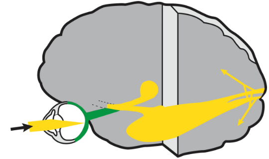
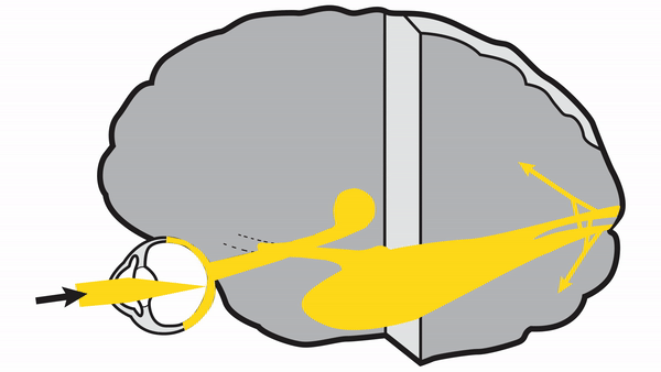
Retina converts optical information into neural signals (“visual transduction”) that travel to the optic nerve
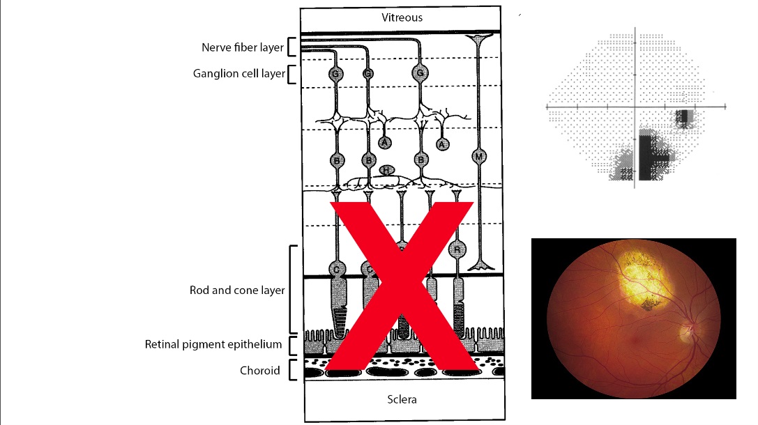
Retinal cones, concentrated mostly in the fovea, mediate high resolution and color vision
Retinal rods, concentrated outside the fovea, gather dim light across broad receptive fields and provide low resolution vision in dim light
Bipolar, horizontal, and amacrine cells receive signals from rods and cones, refine and convey them to retinal ganglion cells
Retinal ganglion cells receive signals from bipolar, horizontal, and amacrine cells and send their axons through the retinal nerve fiber layer to the optic nerve
Retinal nerve fiber layer has three main axon bundles
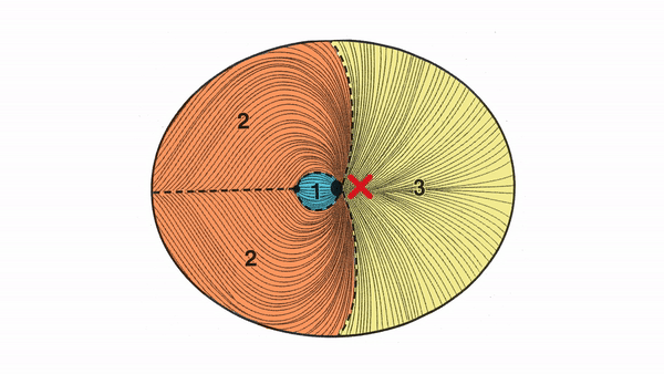
Maculopapillar bundle carries retinal ganglion cell axons from the fovea and retinal region between the fovea and optic disc; mediates high resolution and color signals
Arcuate bundles carry retinal ganglion cell axons from above and below maculopapillar bundle; mediate lower resolution and mostly achromatic signals
Nasal radial bundles carry signals from the nasal retina
Lesions typically cause focal visual field defects called “scotomas”
Outer retinal lesions cause defects whose shape corresponds to the extent of the lesioned retinal area
Retinal ganglion cell and nerve fiber layer lesions cause nerve fiber bundle defects
Maculopapillar bundle lesions produce central scotomas or cecocentral scotomas, mostly from toxic, nutritional, and hereditary conditions
Arcuate bundle lesions produce nasal steps if the lesion is small,
arcuate (scimitar-shaped) defects if the lesion is medium-sized
and altitudinal scotomas if the lesion is large;
these 3 kinds of arcuate bundle defects,
mostly arise from inflammation, compression, ischemia, increased intracranial pressure, or glaucoma
Nasal radial bundle lesions produce temporal wedge defects, mostly from dysplastic optic neuropathies