( of )
Correct: 0
Incorrect: 0
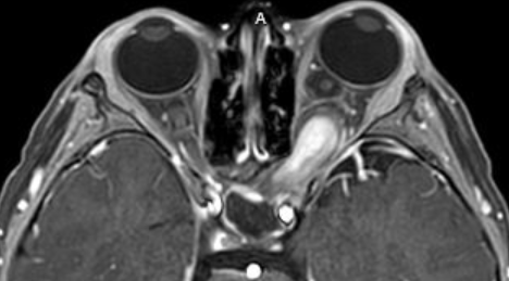
A 5 year old boy is found on optometric examination to have a best-corrected visual acuity of 20/100 (6/30, 0.2) in the left eye and moderate left proptosis. His parents are unaware of when the proptosis occurred and when vision became impaired. The right eye has a normal examination, including formal visual fields. The left eye moves normally, but has an afferent pupil defect and a pale optic disc. Apart from left eye proptosis, there are no other abnormalities on the eye examination and a later neurologic examination is also normal. The MRI, shown here, is interpreted as showing enlargement and enhancement of the intraorbital optic nerve, but no intracranial lesions.
What do you recommend?
Correct!
The imaging features and the patient’s age strongly suggest a diagnosis of pilocytic astrocytoma (“optic glioma”), a World Health Organization Grade 1
(low malignant potential) lesion limited to the orbital segment of the nerve. He may have neurofibromatosis type 1 (NF 1).
There is no evidence that surgery on such a lesion prevents spread to the optic chiasm, hypothalamus, or other optic nerve.
Whether to undertake chemotherapy for a lesion confined to one intra-orbital optic nerve is controversial. Many oncologists would defer because anecdotal evidence of benefit is weak and there are no controlled clinical trials. Current regimens, which are vinca alkaloid and platinum-based, are safe but arduous. There is suggestive evidence that BRAF inhibitors may be more effective. If serial clinical and imaging examinations were to suggest the development of binocular vision loss or imaging involvement of both optic nerves, the optic chiasm or the hypothalamus, chemotherapy would be recommended.
Radiation therapy is not supported by trial evidence and is especially toxic to the developing brain.
Investigation for non-ophthalmic signs of NF 1 is reasonable.
Incorrect
Incorrect