( of )
Correct: 0
Incorrect: 0
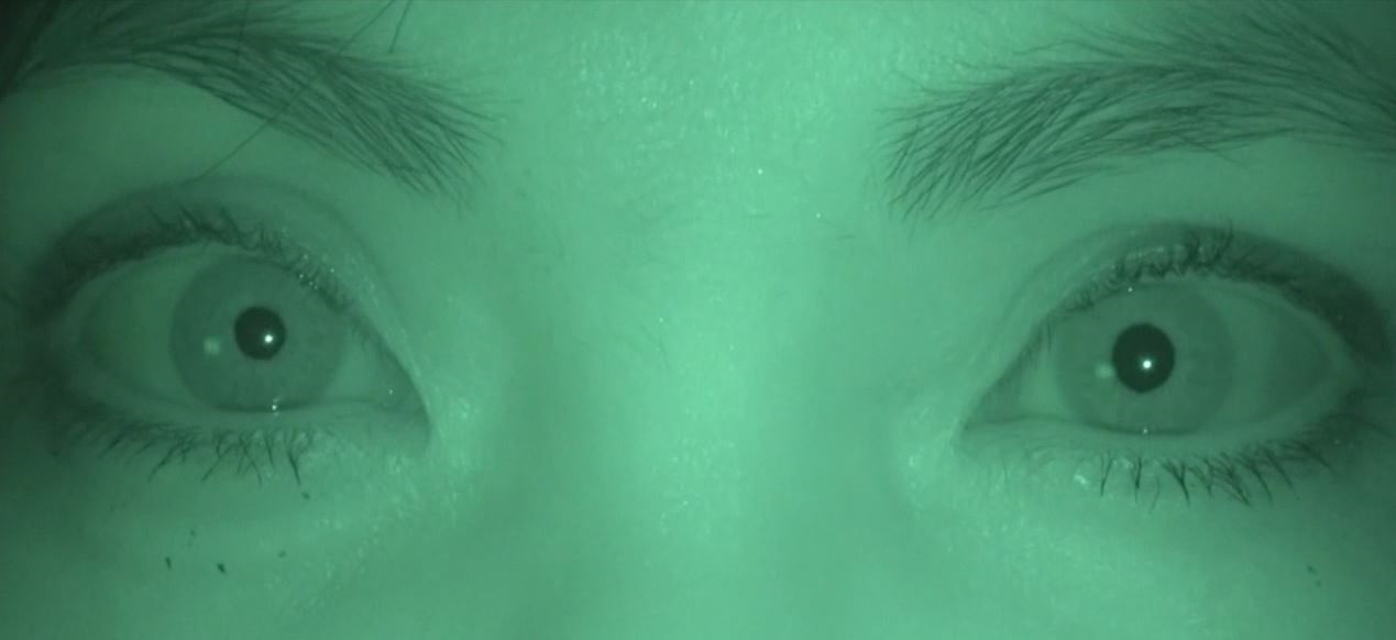
A 54 year old woman is brought to you for anisocoria. As you can see, the left pupil is larger than the right pupil. The right pupil does not constrict to light; the left pupil constricts normally.
Which maneuver should you next perform to help with the diagnosis?
Correct!
Whenever a pupil does not constrict properly to direct light, you should find out whether it constricts to a target placed within reading distance. Why? Because if the
pupil constricts poorly to direct light, but much better to a target placed within reading distance, you have diagnosed “light-near dissociation,” a phenomenon of great
clinical value. In the presence of anisocoria, it signifies a lesion in either the dorsal midbrain/pretectum or the ciliary ganglion/ciliary nerves.
Let us deal first with a dorsal midbrain/pretectum lesion. It causes anisocoria by asymmetric interruption of afferent input to the Edinger-Westphal (parasympathetic) nuclei. It causes light-near dissociation by interrupting the light input but sparing the cerebral input to the Edinger-Westphal nuclei. Other signs of dorsal midbrain syndrome—especially upgaze deficiency—will always be present and they were not noted here. Also, dorsal midbrain lesions do not produce an irregular (oval) pupil or slow (tonic) constriction to a near target, features present here.
The tonic constriction and redilatation you saw here are typical of (Adie) tonic pupil, the result of damage to the ciliary ganglion or ciliary nerves. If you were not sure whether you saw tonicity, you could instill a few drops of dilute (0.1%) pilocarpine in both eyes to search for postganglionic parasympathetic iris sphincter denervation supersensitivity, also a feature of tonic pupil. But as pilocarpine 0.1% is not commercially available, you would have to dilute 1% pilocarpine, a maneuver full of errors and a risk of contamination. Moreover, some patients with preganglionic third nerve lesions will display denervation supersensitivity, misleading you from the correct localization.
You should not be ashamed if you chose, as an answer here, to perform biomicroscopic examination in search of iris defects, including congenital dysplasia or synechiae of old inflammation or trauma. But light-near dissociation would not be present in primary iris sphincter dysfunction.
A small pupil with light-near dissociation would prompt consideration of syphilis-- the Argyll Robertson (AR) pupil. Some people (including this observer) believe that the AR pupil is actually a chronic Adie pupil caused by syphilitic involvement of the ciliary ganglion. You should initiate an evaluation for syphilis ONLY if there are other neurologic signs typical of this disease!
With regard to the other wrong answer choices here: apraclonidine and cocaine are topical agents used when the patient has anisocoria and both pupils constrict normally to light.
In truth, when you are confronted with a patient who has anisocoria and one pupil that does not constrict properly to light, your main job is to make sure that there are no signs of a preganglionic third nerve palsy (ptosis, ductional deficits).