( of )
Correct: 0
Incorrect: 0
A 45 year old woman reports to her optometrist that vision appears “clouded” in her left eye. She is not sure how long that sensation had been present. Visual acuity is best-corrected to 20/20 (6/6, 1.0) in both eyes, but there is an afferent pupil defect in the left eye. The rest of the examination is normal except for these visual fields
Where is the lesion?
Incorrect
Correct!
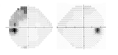
You are looking at a temporal visual field defect in the left eye that has a discrete border aligned to the vertical meridian that passes through fixation.
The visual field in the right eye is normal. The localizing feature here is the configuration of the temporal defect. Because its border is aligned to the
vertical meridian, it signifies a lesion located at the junction of the optic nerve and optic chiasm, where the axons coming from nasal and temporal retina
are separating.
At that junction, axons derived from nasal retina have split off from those derived from temporal retina. Lesions here—no matter their nature—will
preferentially damage the nasal axons to produce this unilateral temporal hemianopic visual field defect. If the lesion were situated farther posterior
toward the optic chiasm, a temporal visual field defect would also be present in the other eye--a bitemporal hemianopia.
This patient had a meningioma growing off the left clinoid process.
Surgical removal led to normalization of the visual fields and disappearance of the afferent pupil defect. Early diagnosis—before the tumor has exerted severe
compression of the optic pathway—improves visual outcome after surgery.
Here is a challenge for you: how could you distinguish an organic from a psychogenic cause of a uniocular temporal hemianopic defect? First, an organic cause would always produce an ipsilateral afferent pupil defect. Second, binocular visual field testing—by confrontation or formal perimetry—will be normal if the defect is organic because the nasal field of one eye covers almost all of the temporal field of the other eye!
This patient had a meningioma growing off the left clinoid process.
Here is a challenge for you: how could you distinguish an organic from a psychogenic cause of a uniocular temporal hemianopic defect? First, an organic cause would always produce an ipsilateral afferent pupil defect. Second, binocular visual field testing—by confrontation or formal perimetry—will be normal if the defect is organic because the nasal field of one eye covers almost all of the temporal field of the other eye!
Incorrect