Parasympathetic Pathway
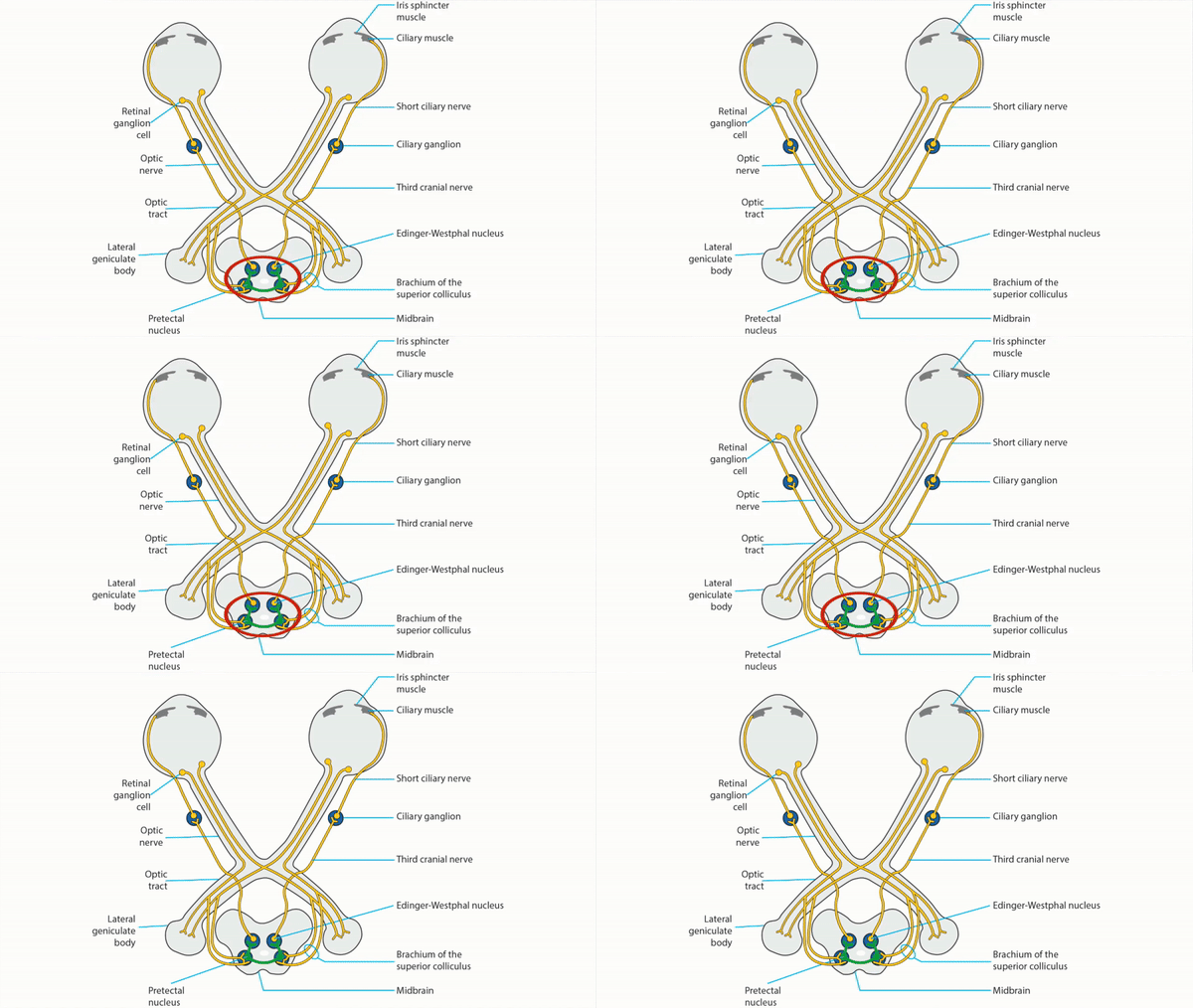
- Light shined in the eye stimulates retinal photoreceptors and intrinsically photosensitive retinal ganglion cells
- Signal travels in the retinal ganglion cell axons through the optic nerves, optic chiasm, and both optic tracts
- Signal branches off in the brachium of the superior colliculus to synapse in the dorsal midbrain pretectal nuclei on both sides
- Axons leave the pretectal nuclei and cross to nuclei on the other side of the dorsal midbrain, so that the afferent signal is distributed equally to both Edinger-Westphal nuclei
- Pre-ganglionic axons from the Edinger-Westphal parasympathetic nuclei carry signals in both third cranial nerves to the ciliary ganglia in both orbits
- Post-ganglionic axons leaving the ciliary ganglia in the short ciliary nerves carry signals to both iris sphincters for pupil constriction and to both ciliary bodies for accommodation


-
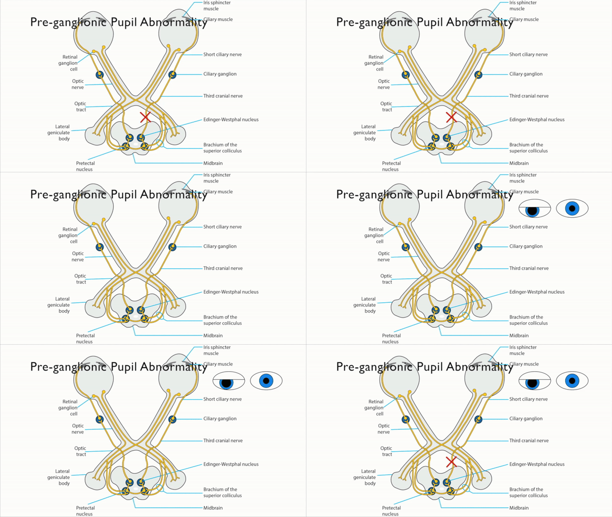
Lesion of one optic nerve
- Produces an afferent pupil defect
- Reduces ipsilateral pupil constriction to light but preserves constriction to a target placed within reading distance because awareness of a near target stimulates a cerebral pathway that bypasses the dorsal midbrain and connects directly to the Edinger-Westphal nuclei (“afferent light-near dissociation”)
- Lesion of both optic nerves
-
Lesion of one optic tract
- Produces a contralateral afferent pupil defect because optic chiasm crossing axons outnumber non-crossing axons
- Produces a contralateral homonymous hemianopia
-
Lesion of one brachium of the superior colliculus
- Produces a contralateral afferent pupil defect
-
Trap: does not cause a visual field defect because a lesion here does not interrupt the visual pathway
- Lesion of the dorsal midbrain
-
Lesion of the third nerve
- Produces ipsilateral mydriasis and impairment of pupil constriction to light, ptosis, and ocular ductional deficits
-
Trap: third nerve lesions never produce mydriasis as an isolated clinical abnormality
- Lesion of the ciliary ganglion or post-ganglionic ciliary nerves


