( of )
Correct: 0
Incorrect: 0
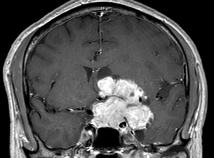
A 15 year old boy reports new headache and impaired vision. Best-corrected visual acuities are 20/200 (6/60, 0.1) in the right eye and 20/400 (6/120, 0.05) in the left eye. There is no afferent pupil defect. All other aspects of the ophthalmic and neurologic examinations are normal. MRI shows this lesion, which was interpreted as showing a craniopharyngioma.
Which of the following statements is true about this lesion?
Correct!
Craniopharyngiomas are made up of congenital cell rests that normally line the throat. These cell rests find their way into the middle cranial fossa and
generate solid and cystic masses.
There are actually two histologic types of craniopharyngiomas: adamantinomatous (occur at any age) and papillary (occur in older adults). Over 90% of the papillary tumors carry BRAF V600E mutations, ushering in a highly effective treatment with inhibitors. There is yet no comparable medical treatment for the adamantinomatous tumors. For them, extensive surgical removal is important, but complete excision is dangerous to vision. High-dose x-irradiation is critical to prevent regrowth, but it works best if much tumor has already been removed. Because these tumors often grow upwards to block the foramen of Monro, they may cause hydrocephalus that often requires ventriculo-peritoneal shunting. In spite of extensive surgery and radiation, cystic regrowth is common and causes rapid loss of vision. The cysts must be approached surgically.
There are actually two histologic types of craniopharyngiomas: adamantinomatous (occur at any age) and papillary (occur in older adults). Over 90% of the papillary tumors carry BRAF V600E mutations, ushering in a highly effective treatment with inhibitors. There is yet no comparable medical treatment for the adamantinomatous tumors. For them, extensive surgical removal is important, but complete excision is dangerous to vision. High-dose x-irradiation is critical to prevent regrowth, but it works best if much tumor has already been removed. Because these tumors often grow upwards to block the foramen of Monro, they may cause hydrocephalus that often requires ventriculo-peritoneal shunting. In spite of extensive surgery and radiation, cystic regrowth is common and causes rapid loss of vision. The cysts must be approached surgically.