( of )
Correct: 0
Incorrect: 0
A 74 year old woman reports being suddenly aware of a haze in the far peripheral field of her left eye. These visual fields were astutely performed on the Goldmann kinetic perimeter after ophthalmoscopy and standard static perimetry had been negative.

Where is the lesion?
Correct!
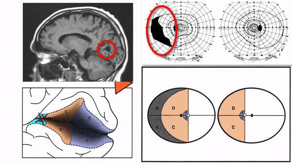 No one would fault you for guessing “retina,” thinking that there was an overlooked abnormality--a retinal inflammation, separation, or scar. But that was not the case here. The
defect derives from the other end of the visual system. Limited to the peripheral temporal field of the left eye, it is easy to miss unless you remember that the temporal field is
normally more extensive than the nasal field. Standard static perimetry, which samples at most the central 30 degrees of the visual field, will always miss this defect. It is called a
“temporal crescent defect,” and it occurs when a lesion—nearly always an infarct--damages the far anterior visual cortex in the opposite cerebral hemisphere
No one would fault you for guessing “retina,” thinking that there was an overlooked abnormality--a retinal inflammation, separation, or scar. But that was not the case here. The
defect derives from the other end of the visual system. Limited to the peripheral temporal field of the left eye, it is easy to miss unless you remember that the temporal field is
normally more extensive than the nasal field. Standard static perimetry, which samples at most the central 30 degrees of the visual field, will always miss this defect. It is called a
“temporal crescent defect,” and it occurs when a lesion—nearly always an infarct--damages the far anterior visual cortex in the opposite cerebral hemisphere
Factoid: this is the only retrochiasmal visual field defect that is not a homonymous hemianopia! By the way, if a lesion can selectively damage the far anterior visual cortex,
it can also selectively spare it. If you play the next video, you will see a “temporal crescent-sparing homonymous hemianopia” caused by a lesion that damaged only the mid-portion
and posterior portion of visual cortex.
Patients who have this defect will be able to see in the periphery of the damaged hemifield and believe that they are safe to drive. Not really!
Incorrect
Incorrect