( of )
Correct: 0
Incorrect: 0
A 25 year old man noticed slowly failing vision in his right eye. He had no pre-existing medical problems. Visual acuity was 20/25 (6/7, 0.9) in the right eye and 20/20 (6/6, 1.0) in the left eye. There was a mild afferent pupil defect in the right eye. The rest of the examination was normal. Here are his visual fields.

Where is the lesion?
Incorrect
Correct!
 This is the most easily recognized visual field abnormality—a bitemporal hemianopia. It is the signature of an extrinsic or intrinsic lesion compressing, inflaming, infarcting, or expanding the optic chiasm.
Actually, the most common cause of chiasmal damage is a mass lesion: pituitary adenoma, meningioma, and aneurysm in adults; craniopharyngioma and pilocytic astrocytoma in children. Why do such lesions produce this defect pattern? Because the crossing axons in the optic chiasm are especially vulnerable to any kind of insult! Patients are slow to notice the defects because the nasal field of each eye almost completely covers the temporal field of the other eye. Although they may eventually sense that peripheral vision is compromised, more often patients present with decreased vision in one eye because the lesion also compromises the function of an optic nerve. Visual fields may show a combination of nerve fiber bundle defects and temporal hemianopic defects (“junction pattern”).
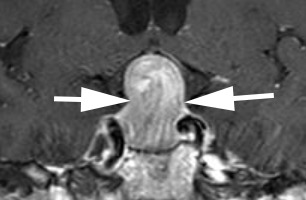
This patient had a pituitary tumor.
The serum prolactin level was normal, eliminating prolactinoma as the type of adenoma. Tip: prolactinomas are treated initially with cabergoline, a dopamine agonist that can dramatically shrink the tumor and restore normal pituitary function, but the medication must be taken indefinitely or the tumor will recur. In this patient, transsphenoidal surgery yielded complete recovery of vision. Early diagnosis of this condition unquestionably improves visual outcome!

Incorrect
Incorrect